Leif Schröder How Can Magnetic Resonance Imaging Be Improved for Early Disease Detection?
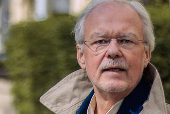
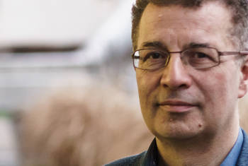
Leif Schröder is a Research Group Leader of the Molecular Imaging Group at the Leibnitz Institute for Molecular Pharmacology in Berlin. Previously, he spent four years at the University of California at Berkeley, partly supported by an Emmy Noether Fellowship from the German Research Foundation, where he worked on new magnetic resonance imaging techniques based on hyperpolarized xenon biosensors. In 2009, he returned to Germany to join the Leibnitz Institute where he led the Starting Grant Project BiosensorImaging funded by the European Research Council until 2015. His research is dedicated to Nuclear Magnetic Resonance (NMR) spectroscopy and imaging where he currently takes a special interest in hyperpolarised biosensors for NMR and Magnetic Resonance Imaging (MRI).
Area of Research
NMR Spectroscopy and Imaging
since 2014
Head of Junior Research Group "Molecular Imaging"
Leibniz Institute for Molecular Pharmacology (FMP) (more details)
Department of Molecular Pharmacology and Cell Biology
2009-2014
Head of the ERC Project BiosensorImaging
Leibniz Institute for Molecular Pharmacology (FMP) (more details)
2009-2009
Head of the Emmy Noether-Group Molecular Imaging
Leibniz Institute for Molecular Pharmacology (FMP) (more details)
2007-2009
Postdoctoral research fellow
Lawrence Berkeley National Laboratory
Materials Sciences Division
2005-2007
Postdoctoral scientist
University of California
Department of Chemistry
2000-2005
Research assistant
Heidelberg University (Ruprecht-Karls-Universität Heidelberg)
Department of Medical Physics in Radiology
2003
Doctor of Natural Sciences
Heidelberg University (Ruprecht-Karls-Universität Heidelberg)
2001
Diploma in Physics
Heidelberg University (Ruprecht-Karls-Universität Heidelberg)
Prizes
- Young Scientist Prize of the International Union for Pure and Applied Physics (2009)
- Dr. Emil Salzer Prize for Cancer Research of the German Cancer Research Center (2008)
- Philips Research Prize for Medical Physics (2005)
Fellowships
- Emmy Noether Fellow of the German Research Foundation (2009)
- Reinhart Koselleck Grant of the German Research Foundation (2016)
- Starting Grant of the European Research Council (2009)
 © FMP
© FMP

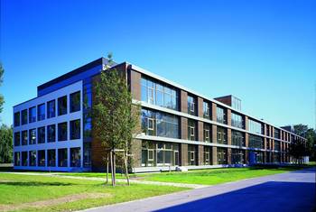
Leibniz Institute for Molecular Pharmacology (FMP)
The FMP conducts basic research in Molecular Pharmacology with the aim to identify novel bioactive molecules and to characterize their interactions with their biological targets in cells or organisms. These compounds are useful tools in basic biomedical research and may be further developed for the treatment, prevention, or diagnosis of disease.
To this aim FMP researchers study key biological processes and corresponding diseases, such as cancer, aging including osteoporosis, or neurodegeneration. They also develop and apply advanced technologies ranging from screening technologies over NMR based methods to proteomics and in vivo models. (Source: FMP)
Department
Department of Molecular Pharmacology and Cell Biology
The focus of research in the Haucke laboratory is the dissection of the molecular mechanisms of endocytosis and endolysosomal membrane dynamics and its role in cell signaling and neurotransmission. The laboratory uses a wide range of technologies that include biochemical and molecular biological approaches in vitro, chemical biology and screening technology, super-resolution and electron microscopy as well as genetic manipulations at the organismic level in vivo. The overarching goal of these studies is to provide a mechanistic understanding of exo-endocytosis and endolysosomal function and its regulation by proteins and lipids and to use this know-how to develop novel strategies for acute chemical and pharmacological interference. (Source)
Map
The technique of Magnetic Resonance Imaging, or short MRI, is a useful and widely used tool in clinical diagnostics. However, the current MRI techniques are not sensitive enough to detect low concentrations of drugs or disease related molecules. LEIF SCHRÖDER explains that MRI is typically based on the detection of water molecules. However, the high water concentration that is always present in the body creates a strong background signal obstructing the signal of dilute molecules so only substances at higher concentrations can be found. In the new approach presented in this video, the researchers used the noble gas Xenon, manipulated its magnetic properties and paired it with a contrast agent which senses specific molecules related to cancer. With this technique, they managed to visualize also molecules in very low concentrations as it is the case for early onset cancer. This approach can help to spot diseases at a very early stage or support drug development.
LT Video Publication DOI: https://doi.org/10.21036/LTPUB10327
Identification, Classification, and Signal Amplification Capabilities of High-turnover Gas Binding Hosts in Ultra-sensitive NMR
- Martin Kunth, Christopher Witte, Andreas Hennig and Leif Schröder
- Chemical Science
- Published in 2015


